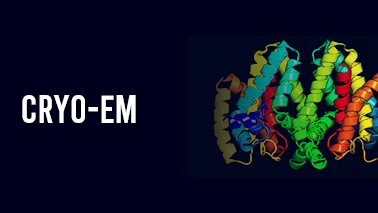
In cryo-EM, researchers rapidly freeze a cell, virus, chemical complex, or other structure to prevent the formation of ice crystals. This maintains the original status of the sample. An electron microscope is used by scientists to blast the frozen sample with an electron beam. This creates a projection of the sample in two dimensions on a digital detector. Scientists build a three-dimensional model of a sample's structure by generating hundreds of projections of the sample from a variety of angles and then averaging these projections. Recent breakthroughs in cryo-EM offer images of proteins and other biological structures, including bigger structures like RNA-protein complexes, in exquisite detail.
Share this page on your timeline
ALSO READ Molecular Biology Structural Biology Folding and Binding Protein Structure Database Protein Engineering Sequence Analysis and Topology 3-D Structure Determination Computational Structural Biology Molecular Modelling and Dynamics Drug Designing and Biomarkers Macromolecular Machines Gene Regulation Cell Signalling Structural Enzymology Structural Bioinformatics Biochemistry and Biophysics Cell Biology Carbohydrate-Protein Interactions and Glycosylation Proteomics and Genomics Structural Biology in Cancer Research Molecular Biology Techniques Advancements in Structural Biology Protein-Nuclic Acid Interactions Membrane Structural Biology Biophysical and Molecular Biological Methods Catalysis and Regulation X-ray Crystallography NMR Spectroscopy cryo-EM Structural Biology in Drug Discovery Structural Biology in Immunology Data Mining in Structural Biology Protein-Protein Interactions Plant Structural Biology Mass Spectrometry in Structural Biology Biomolecular Simulations
Tags
Structural Bioinformatics Conferences
Protein Engineering Conferences
Bioinformatics Conferences
cryo-EM Conferences 2024
Proteomics Conferences
Mass Spectrometry Conferences
Computational Structural Biology Conferences
Protein Chemistry Conferences
X-ray Crystallography Conferences
NMR Spectroscopy Conferences
Cell Signalling Conferences
Computational Structural Biology Conferences 2024